Ask your Dentist if Nanos are right for you! - Part 2: Nanofibers, Nanofilaments and Nanoribbons, oh My!
Investigations into Dental Anesthetics- Part 2
Nanofilaments, Hydrogel, and tiny Nanobots. Follow the red arrow to see the two dancing Nanobots inside the Hydrogel Nanocapsule. (Sample: Lidocaine)
In Part 1 of this investigative microscopy series on Dental Anesthetics, I described a bit about the corporation Septadont and Hydrogel Nanocapsule structures I found. In this post, I will share photos (all 100x or 400x magnification) of a variety of Nanofibers, Nanofilaments and Nanoribbons in the same 8 samples of 5 different Anesthetics:
Lidocaine
Septocaine® Articaine
Mepivacaine Carbocaine®
Marcaine
Prilocaine aka Citanest
‘This way to Transhumanism’ (Sample: Lidocaine)
The truth is: Nanoparticles are used in nanoencapsulation in Anesthetics for drug delivery. For example:
https://www.sciencedirect.com/science/article/abs/pii/S0753332216325458325458
and some of them even have graphene oxide in them:
https://www.sciencedirect.com/science/article/abs/pii/S1011134417313192
https://www.sciencedirect.com/science/article/pii/S0928493119328978#f0045
But what are these Nanofiber, Nanofilament, and Nanoribbon structures doing here?
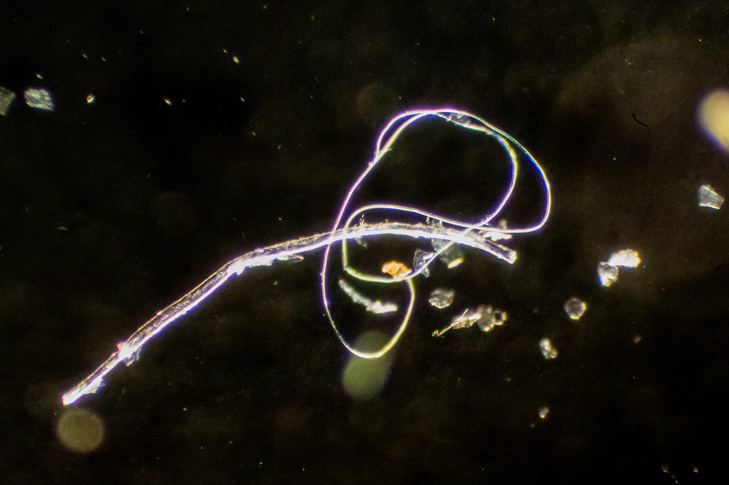
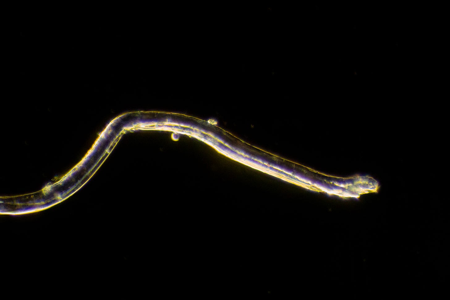
A clear ‘Nanofiber’ and ‘Nanofilament’ under Darkfield; common structures seen these days –in both blood– and virtually everything. (Sample: Articaine)
We believe that these Nanostructures found in the blood, the environment and the food, supplement, medication and water supply are the same, or a more advanced type of self-assembling polymer-based synthetic biology Morgellons fibers that have been found in the environment, as well as coming out of people’s skin. We know from Dr. Ana Mihalcea’s research that the filaments found in the blood in both injected and uninjected people can grow from a nano to a micro scale over time when exposed to EMFs. Indeed, just looking at them on the microscope slide, they get more complex over time as they self-assemble. Homage must be paid to the Carnicom Institute, who first discovered these fibers over 25 years ago. Read oodles of research on the subject and support Clifford Carnicom as well as Dr. Ana’s research here on Substack.
We are working to match up the Nanostructures and processes we find with hundreds of scientific papers, diagrams and patents on Nanotechnology materials to figure our their exact function. I will refer to thinner structures Nanofilaments, the thicker ones with a distinctive tube down the center Nanofibers and the ones that look flattened and fold over Nanoribbons. One paper describes something similar to these filaments as Nanoworms. https://www.researchgate.net/publication/303770433_Polymeric_Filomicelles_and_Nanoworms_Two_Decades_of_Synthesis_and_Application
Why have the formed inside the injection vial? Are some of them just ‘garbage’ Nanomaterials that didn’t fulfill it’s function because it hasn’t yet entered the body? And why are they in virtually everything and everyone now? What we can assume, is that your dentist should not inject any of these synthetic materials into your mouth without your consent.
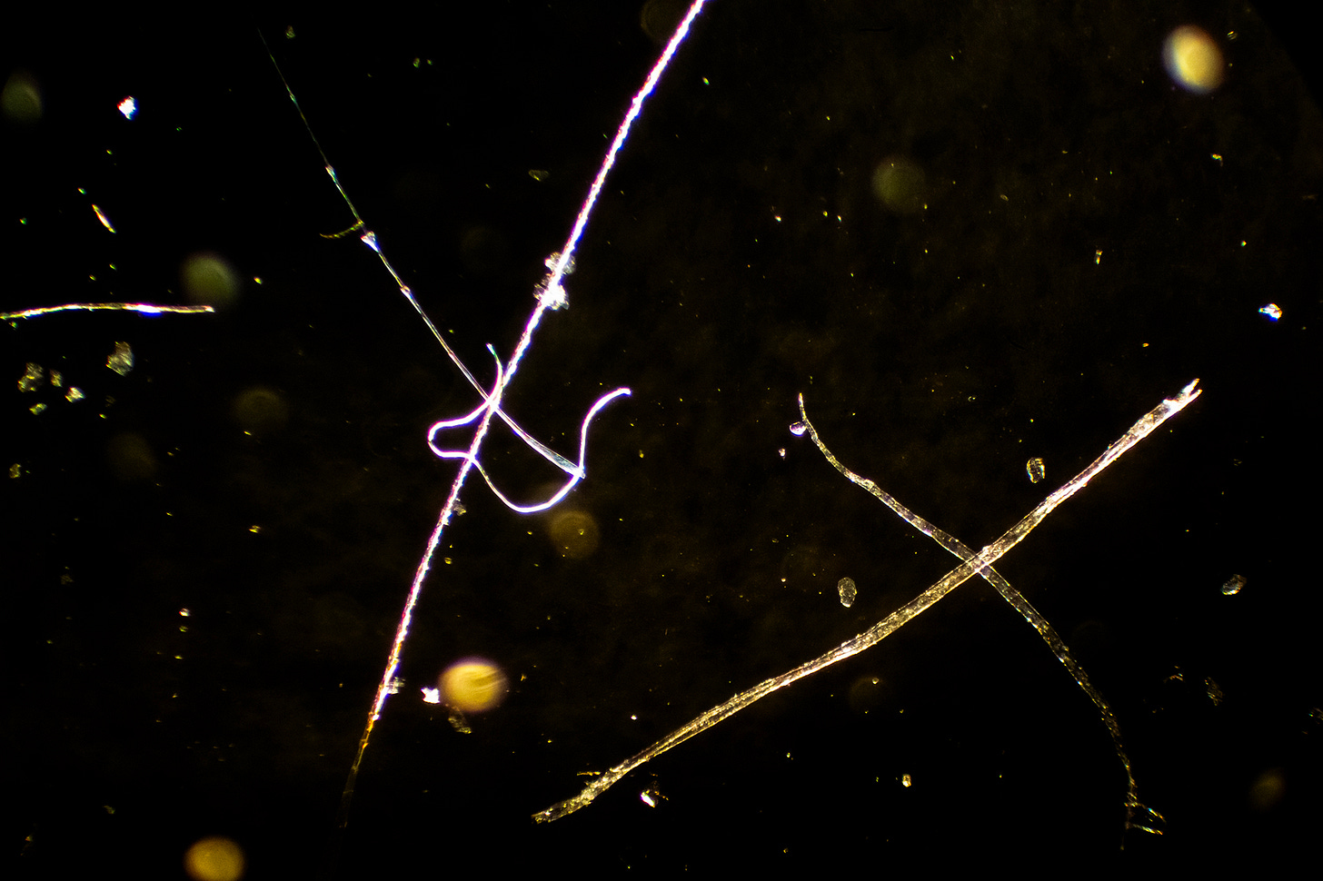
X Marks the Spot! (Sample: Septocaine ® Articaine)
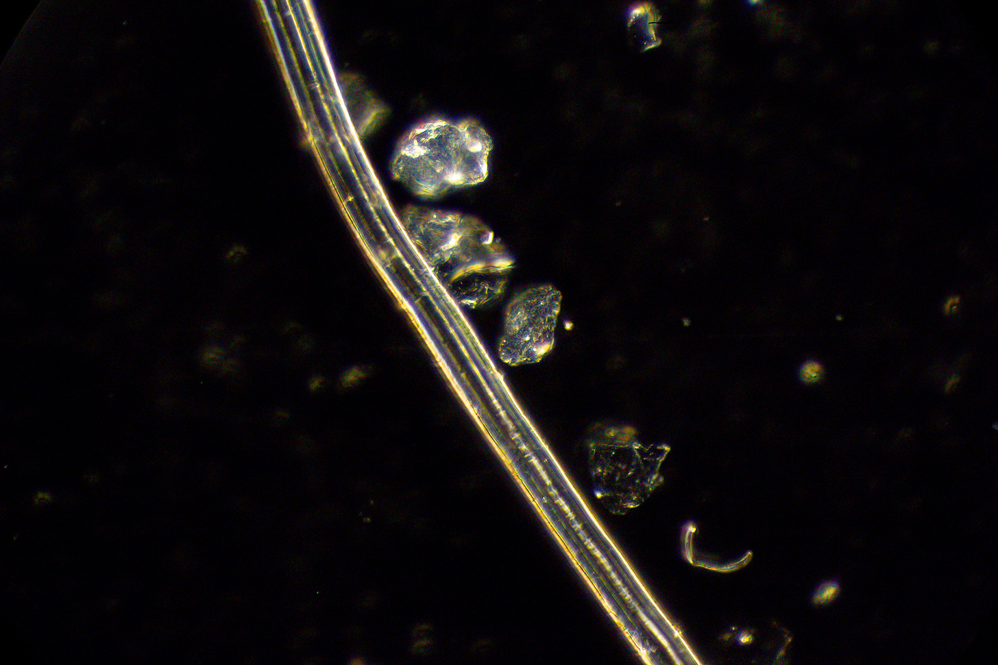
You can see this Nanofiber has a clear center section running through it, indicating a tubular form. (Sample: Mepivacaine/Carbocaine®)
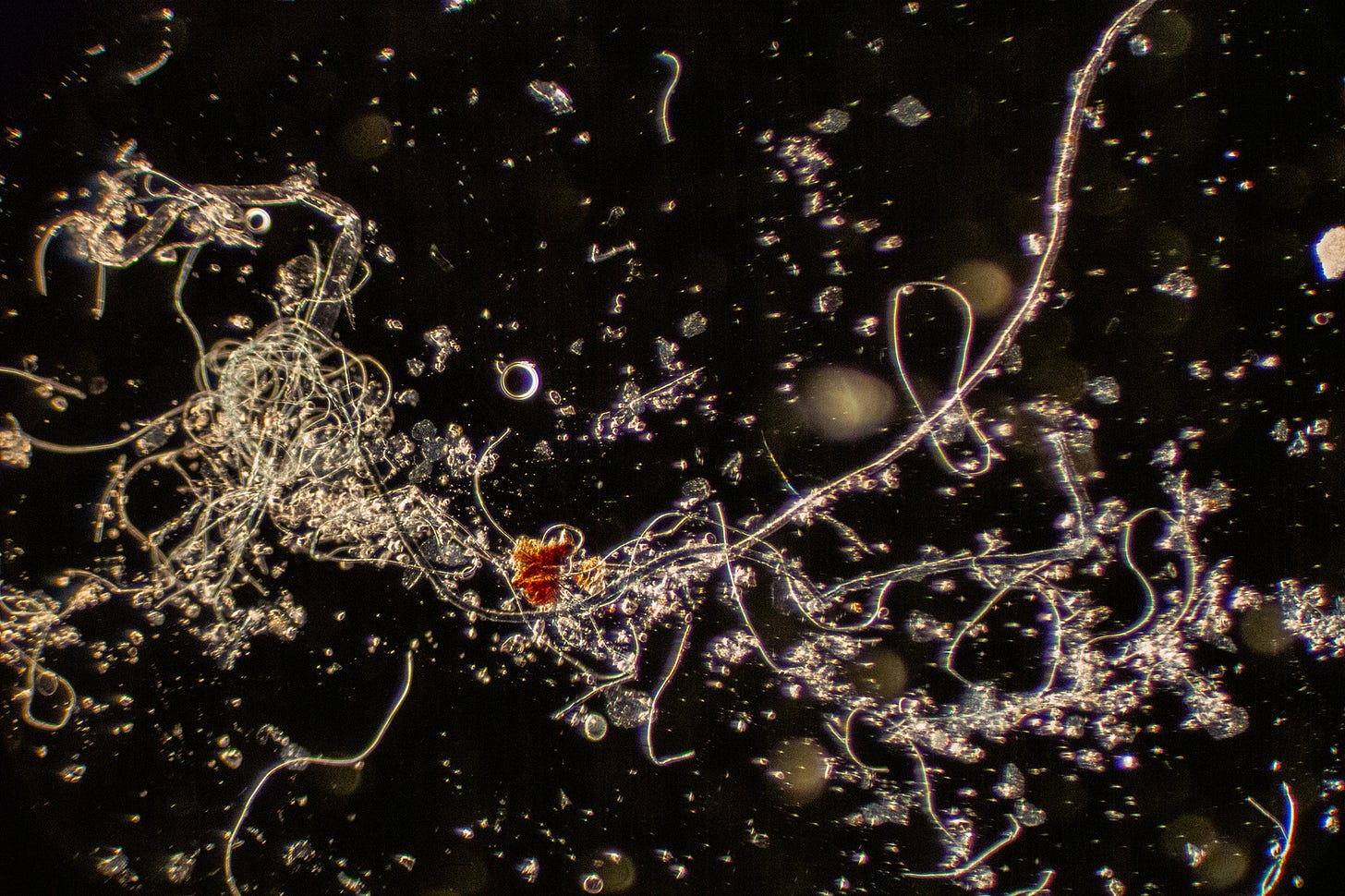
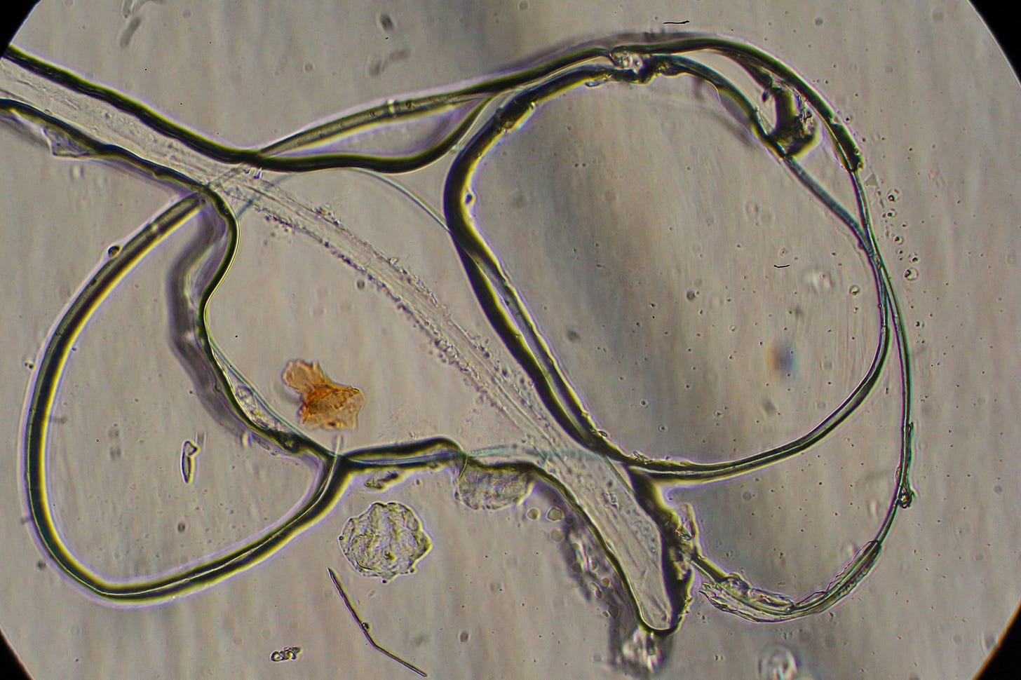
These Nanofibers, Nanofilaments and Nanoribbons can get quite messy. Notice also the two little spherical Nanocapsules, or what I call ‘Nanoportals’, in the upper right quadrant. Not sure what the rusty blob in the center is. I call this one “The Great Nano Garbage Patch of Ocean Microplastics “. (Sample: Septocaine ® Articaine).
This tangled mass of Nanofilaments and Nanofibers I call: “Left over Vermicelli with sun-dried tomato”. (Sample: Septocaine® Articaine).
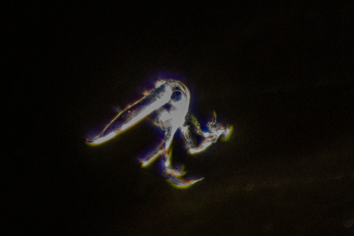
I call this one “Run, Run, Run Away Nano Kangaroo”. (Sample: Citanest/Prilocaine, expired 2020)
Check out this beauty!
I call this photo: “Ghost of Nano Seahorse” (Sample: Marcaine® /Bupivacaine)
A Rainbow of Nanos!
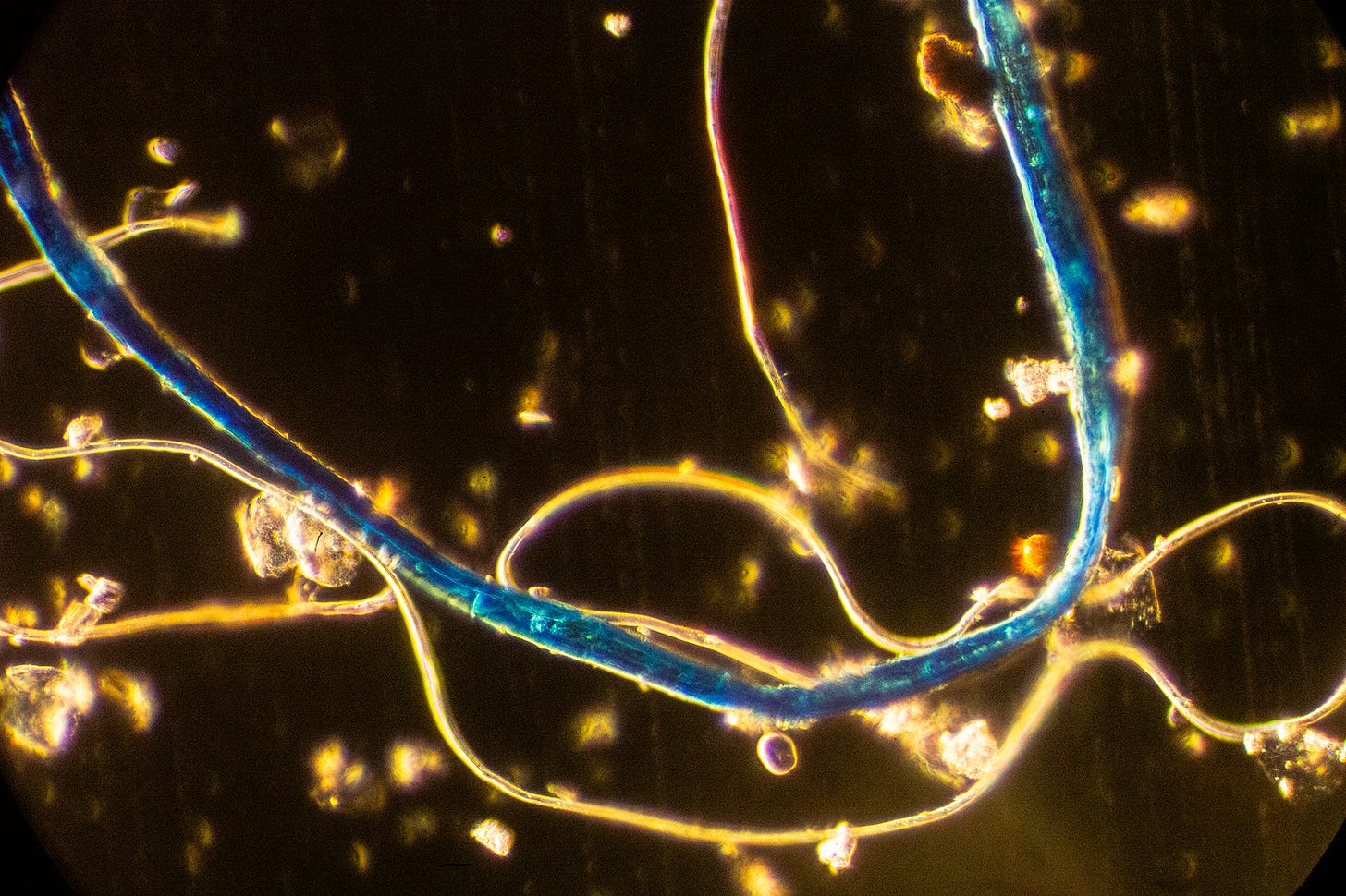
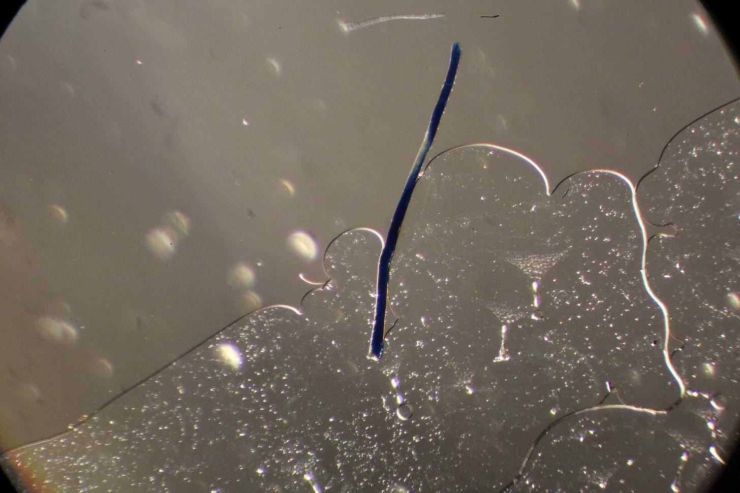
Some Nanos are clear, and others are in colors, such as this bright Blue Ribbon. The others are actually clear, not golden as this appears under Darkfield. I call this one “Bejeweled Nano Necklace”. (Sample: Septocaine® Articaine)
Blue Nanofiber emerging from or being swallowed up by Hydrogel? (Sample: Citanest/Priolocaine, expired 2020)
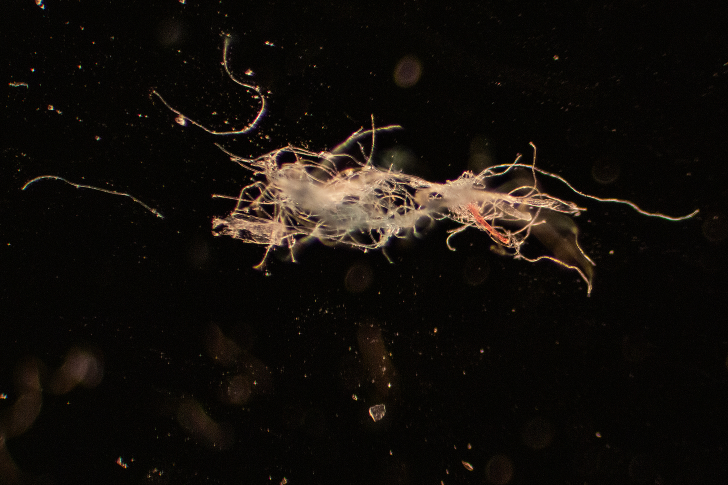
Some are red or pink. This image is in Phase Contrast. Notice the Hydrogel goo surrounding it. (Sample: Septocaine ® Articaine).
Here’s another red “Unidentified Flying Nano” in Mepivacaine Carbocaine®
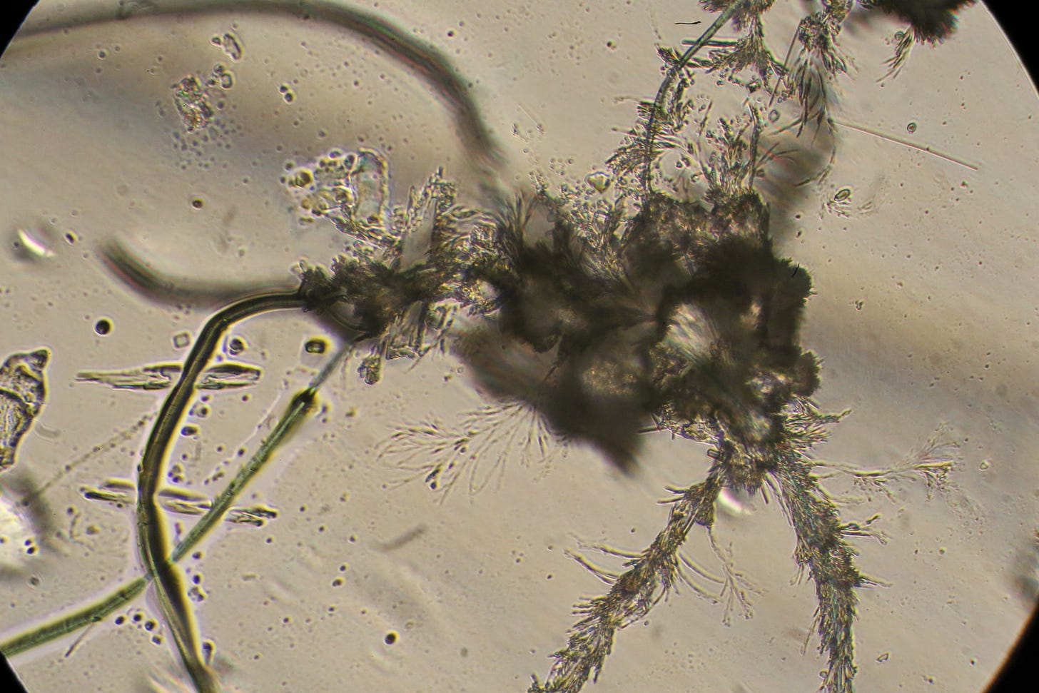
Yum! Check out this nasty nasty dendritic branching formation cluster with a Green Nanofilament on the bottom left. (Sample: Mepivacaine/Carbocaine®) I don’t think this is an example of a synbio creature, with hairy legs, but we don’t really know. I’ve never seen this ink block effect before. I call this one “Creepo Rorschach Nano Test”.
Another Green one with mystery rusty glob. (Sample: Articaine)
Yet another Green one with mystery rusty glob. I call this one “I Took the Wrong Exit on the Spaghetti Nano Junction”. (Sample: Septocaine ® Articaine)
So Cute! “New From Mattel! My Pet Baby Nano Slug: Collect all Four Colors! ” (Sample: Mepivacaine/Carbocaine®)
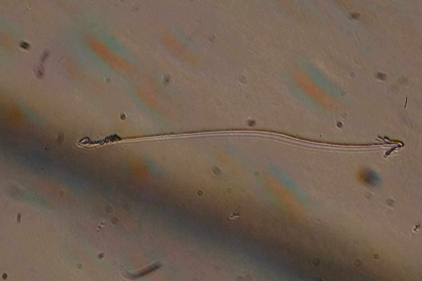
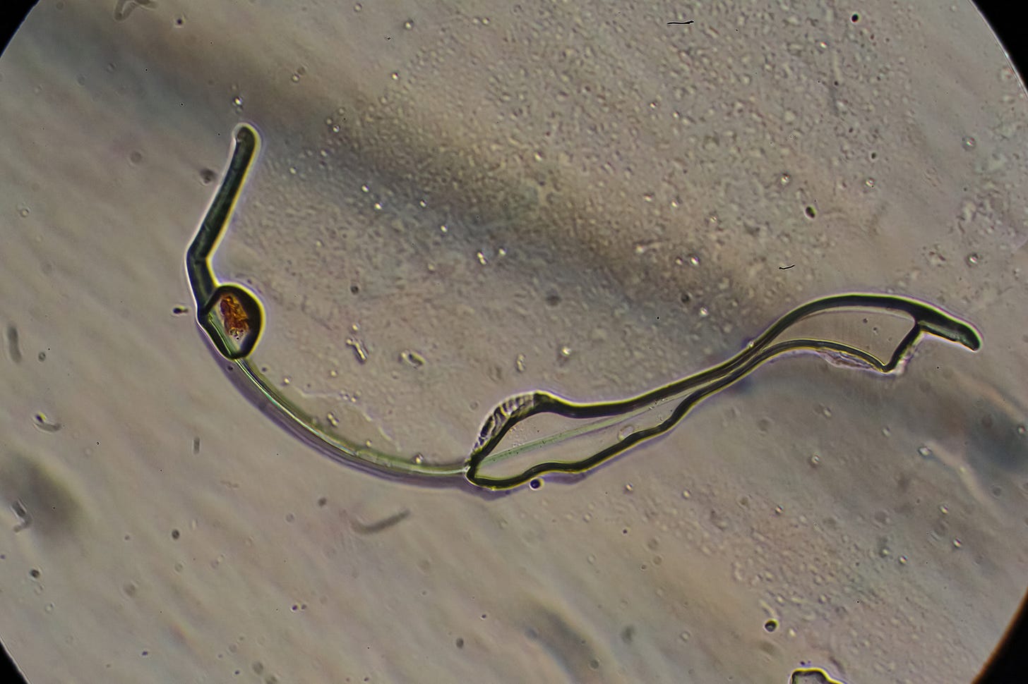
I don’t know what to call this Green Nanofilament with mystery Hydrogel Nanocapsule containing payload, but its nasty. (Sample: Citnest/Prilocaine, expired 2020).
‘Nano synthetic serpents?
OK, these are really scary looking….
“Nasty the Nano Serpent” (Sample: Citanest/Prilocaine, expired 2020)
I call this one, “Nano Nessie, the Lochness Monster”. (Sample: Citanest/Prilocaine, expired 2020)
Caption this Photo! (Sample: Citanest/Prilocaine, expired 2020)
Here is this same little guy swimming though a sea of hydrogel at 100x magnification:
“Weeeeee…..”
OK- So, these ‘Nano serpents’ could be seen as ‘synthetic parasites’ with ‘mouths’ to attach to the inside of your body parts.
Or, are they are like the ‘male’ part of an ‘electrical plug’ that is reaching for a nanosensor ‘chip’, as first captured in Dr. David Nixon’s viral photo of Comirnaty?
Or, could they be Nanoantennas, part of the Internet of Bodies (IoT) and Wireless Body Area Network (WBAN) structure?
Check out this blue one emerging from Hydrogel, found in the blood by a fellow Micronaut, Alien Morphology. Super Nar, dude!
Wait a minute! I know what these things are!
OK, whatever these Nanostructures are, they are part of the synthetic biology/transhumanist agenda, and I really don’t want them in my mouth, so I will be avoiding all dental work that includes injections, until we find an unadulterated brand!
Stay tuned for Part 3!
UN-HACKABLE SWAG!
Meanwhile, if you are an Un-Hackable Animal yourself, consider becoming a Founding Subscriber and receive one of my original Un-Hackable Animal designs embroidered on a baseball cap or knit cap, printed on a T-shirt or a large Bumper Sticker as my Gift to you, or just visit my Etsy shop (Yes, I Know Etsy is evil….working on another storefront!) to purchase for yourself, and find other original anti-transhumanism, pro-human and sovereignty gear that supports my work. Thank You!











Hi! I just discovered your work and I really appreciate the clarity of your images.
I zoomed in on one of them after screenshotting your video and I believe I can see Michaelson or Sagnac interferometer biosensor; a “C-type fiber”(because it looks like the letter, maybe?)
The following link is from the Journal of Biophotonics. It will show you the C-type fiber / Michelson interferometer I am referring to.
https://onlinelibrary.wiley.com/doi/abs/10.1002/jbio.202100068
I hope you don’t mind since this post is public; I’m gonna post a photo of your sample and link to your substack, and the zoomed-in image with an arrow pointing into the middle of the three circles, at the biosensor.
I have pulled a few dozen microscopic devices out of my body and blood and matched them to their patents online.
See my shocking comparisons on my stack!
Nice work.
Image 5 and 6 (after the top video) is near identical to what I have been capturing from the surrounding air/water vapour, inside and outdoors, then viewed under the microscope (brightfield).
You can collect these at your AC Condenser runoff, or inside air filter or dehumidifier water collection. They appear as clear gel sediment at the bottom of any catchment container. Difficult to see at first, but if you stir them about a little they appear black in color.
A very important point is they cannot be captured using a 'desiccant' De-humidifier which simply blows them back into the atmosphere. For De-Humidifier's you can only collect them with a compressor style unit. Desiccant De-humidifier's are taking over as compressor type's appear to being phased out, wonder why?